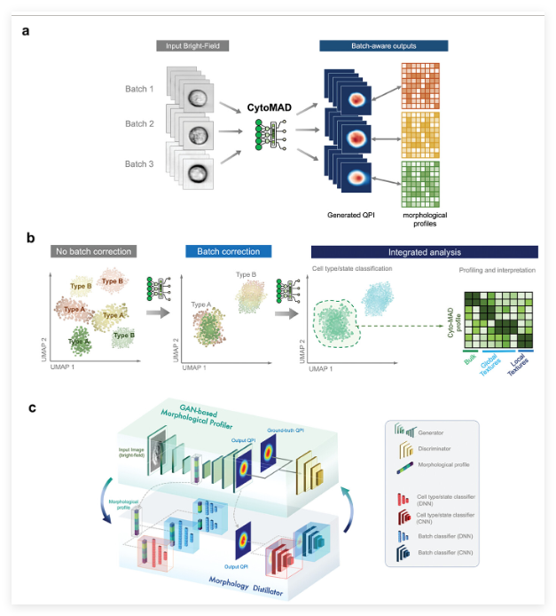
The research team at the University of Hong Kong (HKU) has made breakthrough progress in the field of medical technology recently. They have successfully developed an advanced imaging tool based on artificial intelligence, called "Cell Morphology Against Distillation" (CytoMAD). The innovative technology, led by Professor Qi Kevin from the School of Engineering, aims to achieve single-cell accurate analysis without traditional labeling techniques through a generative artificial intelligence approach, thereby significantly improving the efficiency and accuracy of cancer diagnosis.

CytoMAD technology has demonstrated outstanding performance in collaborative testing at the University of Hong Kong Li Ka-shing Medical School and Mary Hospital, especially in the evaluation of lung cancer patients. By automatically correcting inconsistencies in the imaging process, this technology not only improves the clarity of the image, but also extracts key information that is difficult to detect in the past, providing more reliable data support for medical decisions.
Traditional cell imaging methods usually require staining and labeling of cell samples, a process that is time-consuming and cumbersome. CytoMAD completely changed this situation, eliminating these tedious steps, simplifying the sample preparation process, and greatly speeding up the diagnostic process. The AI model can transform standard bright field images into more detailed representations, revealing cellular properties that are usually difficult to analyze. This transformation is achieved by training generative AI algorithms and can extract cellular mechanical and molecular properties. information.
Currently, many cell imaging techniques rely on slow and expensive processes, which may delay critical treatment decisions. In contrast, CytoMAD provides a tag-free alternative that not only reduces costs but also maintains a high degree of accuracy. By leveraging generative AI, the system converts low-contrast bright-field images into more informative visualizations, deeply analyzing cell morphology without chemical staining.
Another challenge in cell imaging is the variation introduced by the differences between device configuration and imaging protocols, namely the “batch effect”. This inconsistency may hinder accurate interpretation of biology. Many existing machine learning solutions rely on predefined data assumptions, limiting their adaptability. CytoMAD, however, does not require predefined data limitations, allowing for more objective and generalized processing of cellular image analysis.
The advantage of the CytoMAD system is its ultra-high-speed optical imaging technology, which can capture millions of cell images every day. This high-throughput capability accelerates the training, optimization and implementation of AI models. The research team hopes to use this technology to further improve AI-driven biomedical imaging solutions. The ability to quickly process large amounts of cellular data makes CytoMAD a powerful tool in clinical applications and medical research.
In addition to lung cancer diagnosis, CytoMAD may also accelerate drug discovery and reduce the time required for the screening process. The combination of efficient imaging and AI-driven analysis provides a more efficient alternative to traditional methods. Rapid assessment of cell responses to treatments is expected to improve the timeline of drug development and thus bring value to pharmaceutical research.
In the long run, the research team hopes to extend the application of CytoMAD to the predictive medical field, planning to train models to detect early signs of cancer and other diseases. Future developments may focus on integrating the system into clinical practice to enable real-time patient monitoring and personalized treatment planning. AI can analyze massive data and capture subtle cellular changes, which may improve the ability to detect early disease and thus improve the treatment effect of patients.
To drive the study, the team is seeking financial support to track lung cancer patients in a three-year clinical trial to track results using AI-enhanced imaging. This research is expected to promote the wider application of AI in medical diagnosis and improve the efficiency and scalability of medical solutions.
Paper: https://advanced.onlinelibrary.wiley.com/doi/full/10.1002/advs.202307591
Key points:
** The research team has developed CytoMAD, a new AI-driven imaging tool that can improve the accuracy and speed of cancer diagnosis. **
**CytoMAD simplifies the diagnostic process through automatic image correction and analysis. **
** This technology is not only suitable for lung cancer detection, but also accelerates drug discovery and is expected to be applied to a wider predictive medical field in the future. **